- Joined
- Dec 29, 2012
- Messages
- 247
- Reaction score
- 4
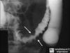
68 yo male patient presents to your clinic with postprandial pain. X-ray below, whats the dx?

That's not it either, though the cells shown in the image can become malignant.
I've made this one super tough; I couldn't answer it if I didn't write it...
hurthle cell is a hashimoto thing. is this just the beginning stages where there's transient thyrotoxicosis?
Well then my attempt to make this tough, just made it a convoluted and poor question...
But now you're on the right track...
Hürthle cell carcinomas account for approximately 3% of all thyroid malignancies and, under the World Health Organization classification, are considered to be a subtype of follicular thyroid cancer. Hürthle cell cancers also are characterized by vascular or capsular invasion and can, therefore, not be diagnosed by FNAB. Tumors contain sheets of eosinophilic cells packed with mitochondria, which are derived from the oxyphilic cells of the thyroid gland. Hürthle cell tumors differ from follicular carcinomas in that they are more often multifocal and bilateral (about 30%), usually do not take up RAI (about 5%), are more likely to metastasize to local nodes (25%) and distant sites, and are associated with a higher mortality rate (about 20% at 10 years). Hence, they are considered to be a separate class of tumors by some groups.
Lol I didn't quite recognize the hurthle cells though
What was the ID for the question? on uworld i wanna look at it
oh ok someone above made a comment that youve been looking too hard at the tough uworld questions lol
Not to belabor the point, but why would you need cytology to diagnose Hashimotos? Through a quick search according to mayo you can get TSH and antibody tests and that should be sufficient to diagnose hashimots. Additionally those cells are showing a certain degree of atypia, which really makes me think it is a neoplasm instead of hashimotos.
Not to belabor the point, but why would you need cytology to diagnose Hashimotos? Through a quick search according to mayo you can get TSH and antibody tests and that should be sufficient to diagnose hashimots. Additionally those cells are showing a certain degree of atypia, which really makes me think it is a neoplasm instead of hashimotos.
Oh you wouldn't need cytology. But I've seen plenty of practice Q's with extraneous (but still relevant) info that's designed to test what you know.
There are plenty of questions that will offer up info that you normally wouldn't have/need before making a Dx...
It was a terrible question though, I thought I was being smart, but I didn't even know that Hürthle cells are associated with other Follicular Thyroid Cancer.
I could probably have gotten away with calling it a diffuse mass rather than just a mass, follicular thyroid cancer is usually noticed as a thyroid nodule rather than a diffusely enlarged thyroid. Additionally, Follicular Thyroid Carcinoma is normally a euthyroid condition rather than a hyper/hypo thryroid condition.
But either way, I was focused on Hürthle cells, and they are not unique to Hashimoto's like I thought they were. That made the question a bad one without a lot more info that I should have included.
Thyroid autoantibodies: Presence of typically anti-TPO (anti-thyroid peroxidase) and anti-Tg (anti-thyroglobulin) antibodies delineates the cause of hypothyroidism as Hashimoto thyroiditis or its variant; however, 10-15% of patients with Hashimoto thyroiditis may be antibody negative
http://emedicine.medscape.com/article/120937-overview

Patient that returns from a mission trip to Nigeria comes in with a red inflamed wrist that is tender palpation, upon examining his eyes you see the above picture.
What do you use to treat this condition?
that's Loa Loa... i think you use Diethylcarbarmazine--something
Patient that returns from a mission trip to Nigeria comes in with a red inflamed wrist that is tender palpation, upon examining his eyes you see the above picture.
What do you use to treat this condition?
pseudotumor cerebri
pseudotumor cerebri


29 year old obese women presents with chronic headaches... no other problems, no medications
6 y/o female brought to physician by mom because she "is too womanly". Bimanual adnexal exam results in a mass on the right.
dx?
Papilledema secondary to obstructive sleep apnea?
Granulosa cell tumor...Call-Exner bodies.
It is papilledema, but it is due to idiopathic intracranial hypertension or pseudomotor cerebri. Obesity predisposes to this condition, although obesity obviously also predisposes to obstructive sleep apnea, but in that case it would be more likely to develop pulmonary hypertension.

Yep, no mention of anything like that to make me thing Pulm-HTN either, I was thinking Obesity--Chronic Headaches--OSA.
Man I hope I don't read too much into questions on the real thing...It's starting to become a habit for me.
I did better on Qbanks before I started reviewing everything.
Yep, no mention of anything like that to make me thing Pulm-HTN either, I was thinking Obesity--Chronic Headaches--OSA.
Man I hope I don't read too much into questions on the real thing...It's starting to become a habit for me.
I did better on Qbanks before I started reviewing everything.
lmao yea. It's always cool when you think for 5 minutes on one question and you narrowed it down to two choices, then when you review, uworld has like 80% picked one choice, and 5% picked the choice you did. And you just sit there like, did people not even spare a thought towards this, yet I spent about 5 minutes on this bs? feelsbadbro

A child from a developing country is seen in the ER for stupor, malaise, and headaches. On physical exam, an enlarged liver is found, and a biopsy is done. Past medical history is significant for palmar erythema, conjunctivitis, and a rash. Here is the biopsy:
What's going on in this patient?
Reye syndrome?
Yea good job lol. I was thinking of leaving some more info out and making it tougher but figured screw it. He had no vaccinations (developing). He got kawasaki->treated with aspirin->infected with VZV for influenza B->reye syndrome with MICROvesicular change (you can tell this just from the histo).

2 different ecg patterns... what might be going on?

Ectopic atrial beat?
Yea good job lol. I was thinking of leaving some more info out and making it tougher but figured screw it. He had no vaccinations (developing). He got kawasaki->treated with aspirin->infected with VZV or influenza B->reye syndrome with MICROvesicular change (you can tell this just from the histo).

alright, who can identify this without a hx?
I am unknowledgable about this, why is microvesicular change important?