- Joined
- Dec 29, 2012
- Messages
- 247
- Reaction score
- 4
68 yo male patient presents to your clinic with postprandial pain. X-ray below, whats the dx?

I think this is would be a problem with the adrenal, while de la chappelle would a problem in the actual sex differentiation. An XX male because one of the Xs has the SRY fragment and they have no MIF so theres no inhibition of development of the mullerian structures
Dude you know this crap... just straight of the dome? that's impressive
Good one! I wonder if this is the same thing as de la chapelle?

will this patient most likely have:
a) diarrhea
b) elevated ESR
c) anemia
d) musculoskeletal pathologies
e) infertility
will this patient most likely have:
a) diarrhea
b) elevated ESR
c) anemia
d) musculoskeletal pathologies
e) infertility
Musculoskeletal Pathologies. Pancoast Tumor.
good. which nerve is most common
That'd be anything on the bottom - lower trunk - ulnar nerve?

That'd be anything on the bottom - lower trunk - ulnar nerve?
heehee I don't actually know. I chose "numbness of 5th digit" on my step so I'm hoping someone here can confirm that ulnar is correct

which side?
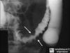
left- sigmoid volvulus.... or the patient might have ingested a giant coffee bean


How would you differentiate this from Hirshprungs?
coffee bean sign
How would you differentiate this from Hirshprungs?
crap, i thought you were jokin earlier... it is a coffee bean sign.
Biopsy would be diagnostic (Hirschsprung has absent inntervation, causing backup). It would also present shortly after birth.
It would also be more like megacolon due to impaction rather than volvulus I'd think.
I think of volvulus as a twisting off of a loop of bowell.
dude, exactly what in the **** is going on here? Are they removing segments of rectal canal and distal colon.... intra-anusly..?management of hirshprung.... looks painful
dude, exactly what in the **** is going on here? Are they removing segments of rectal canal and distal colon.... intra-anusly..?
why is the gapping hole at 1 mo. a good thing? They can poop now but they are totally incontinent?! why not just do a colostomy?
That'd be anything on the bottom - lower trunk - ulnar nerve?
i want to post more but all the pics have the names in the link lol
It's amazing how much book knowledge I've obtained for studying for this test but when used in a practical setting I'm just as likely to diagnose Lyme disease for Endocarditis for SJS for Crohn's if someone doesn't tell me what I'm looking at in words. Good job USMLE
porcelain GB, chronic cystitis?
swallowed pouch. intestinal obstruction

alright, I have a good one.
30 y/o man, recently traveled to south-east asia within the last year where he remembers riding elephants and swimming in local lakes. has a hx of upper right quadrant pain and now presents with noted enlarged liver and spleen along with abdominal fluid wave on abdominal palpation.
Here is a liver biopsy. dx?
33 yo female presents with "a lump in my throat"
ROS positive for: weight loss, increased appetite, and diarrhea.
PE: DTR's 3+ in upper and lower extremities, skin is warm and moist. A mass is palated in her neck lateral to the right and left of the cricoid cartilage.
Biopsy of the mass is performed and is shown in the attached image.
What is the most likely diagnosis?
Edit, wait for the image, I have to resize it...
shistomosomaoa mansoni, is that the portal triad?

damn, that was fast.
apparently, it can be S. Japanoicum or S. Masoni. UW said this is pipestem fibrosis and is pathonomonic for it?
33 yo female presents with "a lump in my throat"
ROS positive for: weight loss, increased appetite, and diarrhea.
PE: DTR's 3+ in upper and lower extremities, skin is warm and moist. A mass is palated in her neck lateral to the right and left of the cricoid cartilage.
Biopsy of the mass is performed and is shown in the attached image.
What is the most likely diagnosis?
Edit, wait for the image, I have to resize it...
im going to guess cancer... and since shes hyperthyroid it must be follicular
That's not it either, though the cells shown in the image can become malignant.
I've made this one super tough; I couldn't answer it if I didn't write it...
sorry, I wikied follicular cell thyroid carcinoma and theres apparently a hurthle cell variant showing the exact same cytology...