- Joined
- Dec 29, 2012
- Messages
- 247
- Reaction score
- 4
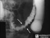
68 yo male patient presents to your clinic with postprandial pain. X-ray below, whats the dx?

ok, this is obviously lung tissue.
hx: 2 weeks ago had an MI. presents now with S3, dyspnea of exertion, and sleeps on top of 3 pillows at night

iron-laden due to chf?
Going to throw this out there but it seems way too easy. Heart Failure cells?
")
ok, this is obviously lung tissue.
hx: 2 weeks ago had an MI. presents now with S3, dyspnea of exertion, and sleeps on top of 3 pillows at night
last one by me for the night...need to go read...but here you guys go...
two different individuals, just providing you two pictures of the same diagnosis


at this stage of the condition...mcc of death would be due to what?

Lichen planus?
last one by me for the night...need to go read...but here you guys go...
two different individuals, just providing you two pictures of the same diagnosis


combo together?
combo together?
blowing systolic murmur, best heard at the apex, and the following skin finding:

Dx?

rheumatic fever (erythema marginatum is shown)
Okay...here's my first contribution
infant/young child with mental ******ation, short stature, and this skin finding:
Hartnup Disease.
That's the Dermatitis of Pellagra right there.
I guess I need to think of a harder oneMiddle-aged adult presents with systolic murmur radiating to carotids and anemia. What's the cause of the anemia?
Mechanical destruction...I think. Does this patient have a mechanical aortic valve, or just really bad stenosis?

A correct answer, but not the one I was thinking of. I'm a bad question-writer
The anemia I was thinking of is coincidental with the cardiac defect (you're right, it's aortic stenosis). There would also be findings upon colonoscopy:
Hopefully that's enough of a hint without giving it away.
edit: didnt see the pic lolA correct answer, but not the one I was thinking of. I'm a bad question-writer
The anemia I was thinking of is coincidental with the cardiac defect (you're right, it's aortic stenosis). There would also be findings upon colonoscopy:
Hopefully that's enough of a hint without giving it away.
Osler-Weber-Rendu
good try but that's not it. This came up in my outpatient IM rotation today and was a PBL case at my school
OK I had to go hunting, but this disease has been cemented in my memory now. I'd never heard of it before.
It's Heyde Syndrome.
aortic stenosis + vWF deficiency/angiodysplasia/iron-deficiency anemia (iirc it's in gunnertraining, but the name isn't listed, just the symptom associations)yep
side question: what's a good short-term pharmacotherapy for this, since the bleeding is due to low vWF (straight out of FA/pathoma)?
Desmopressin.

yep
side question: what's a good short-term pharmacotherapy for this, since the bleeding is due to low vWF (straight out of FA/pathoma)?
Desmopressin.
blowing systolic murmur, best heard at the apex, and the following skin finding:
Dx?


This finding plus this finding
+
equals what syndrome?
This finding plus this finding
+
equals what syndrome?
turcot syndrome?
turcot syndrome?
there's another slightly different syndrome that is the right answer (same family of mutation though). Turcot's would have medulloblastoma or malignant glioma instead of the second pictureCrohn's: cobblestone + erythema nodosum

That's a good guess too, as is Gardner's
clever, but this is erythema nodosum (classically on legs)
It's amazing how much book knowledge I've obtained for studying for this test but when used in a practical setting I'm just as likely to diagnose Lyme disease for Endocarditis for SJS for Crohn's if someone doesn't tell me what I'm looking at in words. Good job USMLE

This finding plus this finding
+
equals what syndrome?
That's a good guess too, as is Gardner's
wow, I have absolutely no idea what that is... damn.27 y/o male just immigrated from Africa shows up with painless ulcerations on his penis that he first noticed 1 week ago. Patient was started on a course of Penicillin G but returned 10 days later when there has been no improvement of his condition.
One of the ulcers is swabbed and the slide below is what is seen in the lab.
What is the arrow pointing to?
What is the disease?
What is the cause of the disease?
Why didn't Penicillin G cure the disease?
(sorry for the Giant picture, especially those of you on phones, but it's a good one and the smaller version lacked)
It's Gardner's right?
FAP + osseous tumors + rentinal hyperplasia?
yes but the mutation is the same in Lynch, not FAP
yes but the mutation is the same in Lynch, not FAP
zzz. you are definitely right tiedyeddog. i thought i was remembering gunnertraining, but i just checked it again and i was wrong
27 y/o male just immigrated from Africa shows up with painless ulcerations on his penis that he first noticed 1 week ago. Patient was started on a course of Penicillin G but returned 10 days later when there has been no improvement of his condition.
One of the ulcers is swabbed and the slide below is what is seen in the lab.
What is the arrow pointing to?
What is the disease?
What is the cause of the disease?
Why didn't Penicillin G cure the disease?