- Joined
- Dec 29, 2012
- Messages
- 247
- Reaction score
- 4
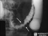
68 yo male patient presents to your clinic with postprandial pain. X-ray below, whats the dx?

yeah...no, i came to the same conclusion and the derm said the same thing too...hopefully it'll go away when the test is done...thanks for the insight...back on topic now
Sure it's the water and not your soap? Also try a water softner, you could be allergic to one of those heavy metals and others aren't.
psoriasis...able to pull the nail off easily
ok to keep with the derm theme

whats this called? and what can it be associated with?
ok to keep with the derm theme
whats this called? and what can it be associated with?
Zinc deficiency? I know that causes nail changes, not sure what they look like though.
Mee's lines, arsenic poisoning?
There's a Beau's line near the tip of the lunula too.
Systemic disease implicated if all 20 nails involved. Pathogenesis: Occur after any severe, sudden, acute, particularly febrile illness; damage to matrix. Etiology: High fever, postnatal, cytotoxic drugs, severe adverse cutaneous drug reaction. Findings: Transverse, bandlike depressions in nail, extending from one lateral edge to the other, affecting all nails at corresponding levels (Fig. 33-23). If duration of disease completely inhibits matrix activity for 714 days, transverse depression results in total division of nail plate (onychomadesis). Multiple parallel lines with chemotherapy. Duration: Thumbnails (lines present for 69 months) and large nails (lines present for up to 2 years) are most reliable markers.
Its Beau's line, not mees though... they look different.
from clinical derm atlas
1. paraesophageal hernia
2. SaO2 = saturation of rbcs with hemoglobin, right? other lung is still working fine and being able to oxygenate the rbcs...so shouldn't it be normal?
1) Defective development of the pleuroperitoneal membrane (which becomes the diaphragm)
2) I gotta think the SaO2 is decreased.
No. Does anyone wanna give it a try before I tell you the answer?
No. Does anyone wanna give it a try before I tell you the answer?
wait what are you talking about?
is this something about how the organs herniate in the posterlateral style into the retroperitoneum by any chance? i remember reading something similar to this in goljan's rr


ok, one more before I peace out for the night:
Patient has recurrent renal colic involving general abdomen pain and recently has had some renal stones.
xray of head, what is the dx?
Paget's?
ok, one more before I peace out for the night:
Patient has recurrent renal colic involving general abdomen pain and recently has had some renal stones.
xray of head, what is the dx?
Nope, but good guess.
ok, one more before I peace out for the night:
Patient has recurrent renal colic involving general abdomen pain and recently has had some renal stones.
xray of head, what is the dx?
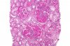
34 year old presents to E.R. with fever, jaundice, and anorexia. Has upper right quad pain.
here is the liver biopsy: What is this type of cell, what does it contain, and what is the disease?
edit: sorry picture is so big. black thing is an arrow pointing to the pathology.
Hyperparathyroidism?
I forgot Paget's presents with normocalcemia since it's a remodeling problem, not a pure osteolysis problem.
Kupffer cells, in alcoholic hepatitis?

that's it, great work.
This is salt and pepper calavarium, which is periosteal thinning.
I am actually not sure how to differentiate this form multiple myeloma.... both would presents with hypercalcemia and bone structural changes...hm. I guess it requires looking for an M spike.
34 year old presents to E.R. with fever, jaundice, and anorexia. Has upper right quad pain.
here is the liver biopsy: What is this type of cell, what does it contain, and what is the disease?
edit: sorry picture is so big. black thing is an arrow pointing to the pathology.
Ito cell aka ? (brain fart atm would get it out of a multiple choice answer though) Contains Vitamin A.

I'll give it up, I think it's a rather obscure pathology but I got a UW question on it.
It's a councilman body, it has acidophilic granules and indicates apoptosis. They are seen are a viral hepatic infection (most likely hep B or C in this case due to drug use). I think they go along with spaces int he liver and hepatocyte balloning.
http://en.wikipedia.org/wiki/Councilman_body
another pic:
arrow is pointing to what? - ive heard 2 different answers, so hopefully somebody can clear it up.
dx?

Those look like the cytoplasmic Ig inclusions of Multiple Myeloma to me.

any idea what they're called...ok so it's Ig inclusions, right? i've heard somebody else saying that their ER accumulations...so was confused
Accumulation of RER product maybe. +1 for the Kip gif
are those neurofibrillary tangles? Intracellular hyperphosphorylated Tau protein, with alzheimer's, chromosome 21. Don't recall how to slow it down though...dx?
chromosomes involved?
treatments to slow down?

are those neurofibrillary tangles? Intracellular hyperphosphorylated Tau protein, with alzheimer's, chromosome 21. Don't recall how to slow it down though...
, right dx of alzheimer's...but that's Abeta amyloid...didnt know what it was because i've always seen questions with this type of pictureI can't figure out how to post images, too lazy to save as.
lol. You can directly link to ones you get online without saving them if you want
Okay I'll contribute to this since I don't want it to die out. Dx?
It couldn't be diffuse proliferative glomerulonephritis could it?
It couldn't be diffuse proliferative glomerulonephritis could it?
im going to go with nodular glomerulosclerosis

Hah, didn't realize the file name had the description. Yeah its diffuse proliferative lupus neprhitis, aka diffuse proliferative glomerulonephritis.yep, looks like a high mag image of diffuse proliferative lupus nephritis to me


newborn. ddx?