As I sit here leafing through my texts, I see sources pointing in both directions.
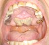
Nanda's "Biomechanics and Esthetic Strategies in Clinical Orthodontics" says that symphyseal DO results in both basal and alveolar bone being formed. It states that case selection is important and the ideal patient for symphyseal DO would have either a constricted maxilla and bilateral buccal crossbites in the dentition, or a constricted maxilla and mandible where you want to widen both to alleviate crowding. It concludes that this procedure is basically not performed as frequently right now, but as more people do it, more data will be produced.
Proffit's "Contemporary Orthodontics" says that orthodontic expansion only at the alveolar bone level to correct mandibular crowding has doubtful stability, especially if you expand the canines without retracting them. But then it gets wishy-washy about whether basal bone expansion with symphyseal DO would be a more stable choice. Instead the text seems to suggest that the orthodontist should just skip either attempt at widening the mandibular bone to alleviate crowding and just treatment plan the extraction of mandibular premolars instead. Then it implies that the lip and cheek pressure against the mandibular canines expanded with symphyseal DO might be an issue as well.
So there isn't much data out there to support basal bone expansion via symphyseal DO. Looks like Guerrero is the one who has done some studies on it since he is credited with doing it with a tooth-borne hyrax device. There seems to be a growing anti-extraction sentiment in ortho, so I'm just trying to explore all options out there to alleviate crowding. This is why I'm wondering how often you OMS residents encounter symphyseal DO in your residencies or know of it being done in private practice.