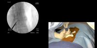
New Vertos/MILD Study: Game changer?

This forum made possible through the generous support of SDN members, donors, and sponsors. Thank you.

drusso

Full Member
Moderator Emeritus
Lifetime Donor
Joined
Nov 21, 1998
Messages
12,586
Reaction score
7,005

New Study Demonstrates 5-Year, Long-Term Durability​

The investigators at the Cleveland Clinic recently published an additional independent, retrospective cohort study which further explores the long-term durability of mild® at 5 years. They followed LSS patients receiving the mild® Procedure at the Cleveland Clinic between 2010 and 2015.

The recently published paper, The Durability of Minimally Invasive Lumbar Decompression Procedure in Patients with Symptomatic Lumbar Spinal Stenosis: Long Term Follow-Up, explores how many patients were able to avoid surgical decompression after the mild® Procedure over a five-year period. Changes in pain level using the Numeric Rating Scale (NRS) and opioid medication utilization using Morphine Milligram Equivalent (MME) dose per day from baseline to 3-, 6-, and 12-months post-mild® Procedure were also collected, along with post-procedure complications.

mild® is a minimally invasive decompression procedure that addresses a major root cause of patients’ stenosis without having to undergo an invasive open surgery or leave implants behind. It has the safety profile equivalent to an epidural steroid injection (ESI), but with lasting results and is often compatible for patients with existing risk factors that, traditionally, may not be candidates for open surgery.1 According to the study, the mild® Procedure significantly decreased the incidence of surgical decompression at the same treatment level(s) as mild® intervention during five-year follow-up. Nine out of 75 patients required lumbar surgical decompression at the same level during the follow-up period, making the annual incidence of same-level lumbar-decompression surgery just 2.4%. Subjects experienced statistically significant pain relief and reduction of opioid medications utilization at 3, 6, and 12 months compared to baseline. There were no major complications reported.

Members don't see this ad.
 
my personal experience with MILD - mediocre result at best.
if anyone else has great outcome and clinical pearls to share let me know. i personally do not like the radiation and vertiflex works better in my experience
 
  • Like
Reactions: 1 users
Members don't see this ad :)
my personal experience with MILD - mediocre result at best.
if anyone else has great outcome and clinical pearls to share let me know. i personally do not like the radiation and vertiflex works better in my experience
how is the radiation any worse than that with SCS?

6 years to complete the study after finished collecting data.
last patient was enrolled in 2015.

followed for 5 years, which would be 2020.... so really 1 year out.


study is not really a study but data analysis of patients who had MILD.

as with all interventional treatments, will be difficult to perform any randomized studies.

 
  • Like
Reactions: 1 user
how is the radiation any worse than that with SCS?


last patient was enrolled in 2015.

followed for 5 years, which would be 2020.... so really 1 year out.


study is not really a study but data analysis of patients who had MILD.

as with all interventional treatments, will be difficult to perform any randomized studies.

Haven’t tried the new technique but I was usually at 5+ minutes of fluoro for a MILD. SCS is usually less than a minute for me.
 
really... I'm getting 2 1/2 minutes, even on multiple levels. my 2 lead SCS trials are typically about 2 minutes.
 
  • Like
Reactions: 2 users
Just like everything we do, we use more fluoro and then use less as we go on and become more proficient at it.
 
I just did an 11 min trial. Implant is today.
Screen Shot 2021-04-23 at 4.56.45 PM (1).PNG
 
  • Like
Reactions: 1 user
Wow. How/where did you do a trial on that? Axial pain?
 
Wow. Ballsy
I’ll have to send similar ones to you. I’m not brave enough to drive an SCS lead while creating significant leg pain from cord pressure.
 
Wow. Ballsy
I’ll have to send similar ones to you. I’m not brave enough to drive an SCS lead while creating significant leg pain from cord pressure.
Implant today. Strangely, it took just under an hour. Average for me. No issues getting in or up. No wet tap. To steering issues. I cannot predict what cases will go tough. n=lots
 
  • Like
Reactions: 1 user
Members don't see this ad :)
If you’re not good at epidurograms during the MILd procedure you will NOT get good results .... Vertos has taken the epidurogram out of the instructions because so many cant do it or find it tedious having to manage multiple insertional needles . Imo you identifying the posterior elements much better, you decompress the HLF much better and with more confidence (advance deeper). Finally you can see the repeat epidurogram remodel/expand in vivo, which again IMo gives you a better indication of your decompression.

Also, you need to select the right patient : ie neurogenic Claudication NOT a radiculopathy secondary to lateral foraminal stenosis , which is out of our pay grade currently ...
 
  • Like
Reactions: 1 users
If you’re not good at epidurograms during the MILd procedure you will NOT get good results .... Vertos has taken the epidurogram out of the instructions because so many cant do it or find it tedious having to manage multiple insertional needles . Imo you identifying the posterior elements much better, you decompress the HLF much better and with more confidence (advance deeper). Finally you can see the repeat epidurogram remodel/expand in vivo, which again IMo gives you a better indication of your decompression.

Also, you need to select the right patient : ie neurogenic Claudication NOT a radiculopathy secondary to lateral foraminal stenosis , which is out of our pay grade currently ...

Planning to do the training in next couple months. What's the difficulty with epidurogram? Is it significantly different from our usual interlaminar esi?
 
  • Like
Reactions: 1 user
Planning to do the training in next couple months. What's the difficulty with epidurogram? Is it significantly different from our usual interlaminar esi?
no it's not. just in a tight space so higher chance of dural puncture. i generally use a small gauge tuohy (22 ) for it.
epidurogram combined with continuous fluoro during laminotomy/ligamentum decompression boosts my fluoro time into stratosphere. i try with pulsed/low dose/focused beam but still the time ends up being quite long for me.
 
  • Like
Reactions: 1 user
If you’re not good at epidurograms during the MILd procedure you will NOT get good results .... Vertos has taken the epidurogram out of the instructions because so many cant do it or find it tedious having to manage multiple insertional needles . Imo you identifying the posterior elements much better, you decompress the HLF much better and with more confidence (advance deeper). Finally you can see the repeat epidurogram remodel/expand in vivo, which again IMo gives you a better indication of your decompression.

Also, you need to select the right patient : ie neurogenic Claudication NOT a radiculopathy secondary to lateral foraminal stenosis , which is out of our pay grade currently ...
I actually saw an ideal pt for Mild yesterday. 94, neurogenic claudication, nearly perfect discs, 2 level stenosis centrally caused by a 2.4mm thick LF. I was going to train for it, but my new practice doesn't really do hospital based cases. I'm trying to decide if I want to go ahead and learn it
 
  • Like
Reactions: 1 user
Planning to do the training in next couple months. What's the difficulty with epidurogram? Is it significantly different from our usual interlaminar esi?
It’s like perc spinal stimulator implants . You need to manage the epidural needle, the 12-10g introducer trochar, bone and tissue sculptors ( I don’t use the plastic mound on the skin) and going back and forth between ap, clo views. If you are proficient in scs stim, kyphos, you should be fine . I just think for your first 30 cases do the epidurogram for anterior safety margins , and confidence that you have an expansion in the epidurogram at the level your decompressing...
 
  • Like
Reactions: 1 user
I actually saw an ideal pt for Mild yesterday. 94, neurogenic claudication, nearly perfect discs, 2 level stenosis centrally caused by a 2.4mm thick LF. I was going to train for it, but my new practice doesn't really do hospital based cases. I'm trying to decide if I want to go ahead and learn it
Do these have to be done at a hospital? Or can they be done at an asc?
 
I actually saw an ideal pt for Mild yesterday. 94, neurogenic claudication, nearly perfect discs, 2 level stenosis centrally caused by a 2.4mm thick LF. I was going to train for it, but my new practice doesn't really do hospital based cases. I'm trying to decide if I want to go ahead and learn it
i do them in the ASC.

at the ASC i go to, they automatcially get MAC anesthesia, but i wonder if it is absolutely necessary.

imo, a ton easier than an implant. and the likelihood of a complication is much less.

really, imo all you have to be comfortable with is CLO imaging. results with and without epidurogram apparently no different, but i dont have the data. ive done with both. i do like using the epidurogram only because you can give them a dose of steroids before you remove needle. it may be giving a false sense of accomplishment, though. there greater risk of wet tap from the epidurogram than the actual procedure.

if you are worried about safety, just use the rongeur, dont use the tissue sculptor, and dont get an epidurogram. the rongeur is like a large Tuohy - ie a blunt edge.
 
  • Like
Reactions: 1 user
i do them in the ASC.

at the ASC i go to, they automatcially get MAC anesthesia, but i wonder if it is absolutely necessary.

imo, a ton easier than an implant. and the likelihood of a complication is much less.

really, imo all you have to be comfortable with is CLO imaging. results with and without epidurogram apparently no different, but i dont have the data. ive done with both. i do like using the epidurogram only because you can give them a dose of steroids before you remove needle. it may be giving a false sense of accomplishment, though. there greater risk of wet tap from the epidurogram than the actual procedure.

if you are worried about safety, just use the rongeur, dont use the tissue sculptor, and dont get an epidurogram. the rongeur is like a large Tuohy - ie a blunt edge.
I don’t disagree here. However our procedure is percutaneous and not open. We need a metric to assess our debridement . I guess you can guestimAte your decompression by eye balling your tissue removal. You’d have to memorize the MRI and assume a lot of factors. A more rational approach is to repeat the epidurogram after your bilateral or unilateral decompression. It was originally recommended and conceptualized. Again my experience, technique is still developing .
 
Please help me understand something about the epidurogram for mild. I understand it delineates the dorsal epidural space for procedural safety and depth one can work to in ligament. With regards to the post mild epidurogram, in terms of showing better flow, how is this assessed/quantified? Ive done many thousand esi, as have most others on here, and I can’t say I see distinct reliable different patterns/flow when I inject in a stenotic canal vs patent canal ie with small disc herniation. I could conceptualize more flow on a myelogram post decompression, but don’t quite understand seeing reliably better epidural contrast flow. I Inject stenotic spines all the time and see fairly localized flow 1-2 levels up and/or down in some and multiple more levels in others without clear rhyme/reason. Same in c spine. Only occasionally do I see what I would describe as constricted/narrow flow at level of central/lateral recess stenosis. Some severely stenotic levels still have a thick stripe of epi fat on T1 mri. I think pattern seen is more a function of location of contrast in epidural space on clo, such as in the Gill articles. Granted, perhaps the contrast volume on mild is higher, but I’m just failing to see how this reliably shows decompression. Please educate me. Thanks.
 
  • Like
Reactions: 1 users
you eyeball it and it makes you feel better.

in all honesty, the one person that did not get better was one of the people that I swear had more flow post procedure. N about 10.
 
  • Like
Reactions: 1 users
Please help me understand something about the epidurogram for mild. I understand it delineates the dorsal epidural space for procedural safety and depth one can work to in ligament. With regards to the post mild epidurogram, in terms of showing better flow, how is this assessed/quantified? Ive done many thousand esi, as have most others on here, and I can’t say I see distinct reliable different patterns/flow when I inject in a stenotic canal vs patent canal ie with small disc herniation. I could conceptualize more flow on a myelogram post decompression, but don’t quite understand seeing reliably better epidural contrast flow. I Inject stenotic spines all the time and see fairly localized flow 1-2 levels up and/or down in some and multiple more levels in others without clear rhyme/reason. Same in c spine. Only occasionally do I see what I would describe as constricted/narrow flow at level of central/lateral recess stenosis. Some severely stenotic levels still have a thick stripe of epi fat on T1 mri. I think pattern seen is more a function of location of contrast in epidural space on clo, such as in the Gill articles. Granted, perhaps the contrast volume on mild is higher, but I’m just failing to see how this reliably shows decompression. Please educate me. Thanks.
There are advanced peer seminars and webinars if you are interested.
I like multiple metrics to determine my decompression vs just eyeballing it or basing on tissue debridement per patient.
This is from MILD:
  • The epidurogram may have a “scalloped” appearance, representing ligamentum flavum enlargement.
  • The reduction in the “scalloped” shape, or a thickening and straightening of the epidurogram, is expected as decompression of the ligamentum flavum tissue occurs.
  • Only a small amount of contrast is required to obtain the necessary epidurogram (typically <1 cc). Intraoperative replenishing or “blushing” of the epidurogram (using <0.5 cc of contrast) may be needed for optimum visualization (Figures 38-5and 38-6).
 
  • Like
Reactions: 1 user
There are advanced peer seminars and webinars if you are interested.
I like multiple metrics to determine my decompression vs just eyeballing it or basing on tissue debridement per patient.
This is from MILD:
  • The epidurogram may have a “scalloped” appearance, representing ligamentum flavum enlargement.
  • The reduction in the “scalloped” shape, or a thickening and straightening of the epidurogram, is expected as decompression of the ligamentum flavum tissue occurs.
  • Only a small amount of contrast is required to obtain the necessary epidurogram (typically <1 cc). Intraoperative replenishing or “blushing” of the epidurogram (using <0.5 cc of contrast) may be needed for optimum visualization (Figures 38-5and 38-6).
Thanks. I was not thinking of the pattern in clo. That makes sense as I do routinely see that when place needle through lfh on clo on an interlam. This is not a procedure I’ve trained on or intend to use.... more so a curiosity on my part. I’d still refer my parents for an endoscopic decompression by an experienced MIS surgeon, but I do see a place for this in a limited patient population.
 
This just popped up in my email. Recommend attending? It’s $5000 buckaroos but I don’t see how else I would learn it.
 

Attachments

  • 193C21BC-CB20-4751-AC1D-DD6FCDA4A16A.jpeg
    193C21BC-CB20-4751-AC1D-DD6FCDA4A16A.jpeg
    526.4 KB · Views: 118
  • 5D3653D7-4EE4-4DA0-ABD2-5D2C30464B7F.jpeg
    5D3653D7-4EE4-4DA0-ABD2-5D2C30464B7F.jpeg
    167.2 KB · Views: 110
NO!

call your rep.

there are online Zoom courses that are free of charge from the company. Hour long.

I also played with the model that the rep had.

then shadowed a couple of procedures at a local ASC.

I spent 0$. Well maybe a few bucks on buying coffee for shadowing...
 
  • Like
Reactions: 1 user
NO!

call your rep.

there are online Zoom courses that are free of charge from the company. Hour long.

I also played with the model that the rep had.

then shadowed a couple of procedures at a local ASC.

I spent 0$. Well maybe a few bucks on buying coffee for shadowing...
Are you talking MILD or the endoscopic surgery?
 
I think MILD can work very well - and why wouldn't it. It frees up the ligament to be more pliable.

My problem - is it takes me 2+ hours to do a case (i've only done a few). A surgeon can decompress that single level in 30 minutes - and is much more definitive.

My other problem - the company has never been honest about real complications.
 
This just popped up in my email. Recommend attending? It’s $5000 buckaroos but I don’t see how else I would learn it.
Considering how few insurances cover this and the cost of the equipment to actually do these cases, I guess the question would be how long would it take you to make back your money.

unless you do a lot of PI , or have a unique insurance situation, I don’t understand such a course is a good value.
 
Reviving MILD thread. I listened to their webinar and planning my first case next week. I took some screenshots of their video. Some of the pics seems awfully close/violating the VILL. I assume their positioning is more lateral on this CLO view and probably ok, but I would not be comfortable going that far. After watching the video and doing the demonstration my thoughts/questions are as follows:

1) Should I do epidurogram AT the level vs above? I'd prefer at the level. Seeing contrast at the level would make me much more comfortable regarding safety backstop, but risk of dural puncture at stenotic level. The rep recommends above the level or not at all. Above the level seems like it may not spread inferiorly much of the time.
2) Use midline 'streamlined' technique rather than two incision technique
3) use MAC. Colleagues here starting have done all GA. Big advantage I see for this vs surgical decompression seems to be MAC. Is it fine under true MAC (e.g. I can talk to the patient)? Do you make multiple passes with a local needle to superior and inferior lamina or one seems to be ok?
4) With both the bone ronjeur and tissue sculpter, it seems you are pushing the trocar in a medial lateral direction as you take bites. Is that right?
5) And last I suppose any thoughts on these screenshots? That is too far for my comfort.

Tagging @Ducttape and @10KHertz in case you have more pearls. Thanks
 

Attachments

  • mild1.jpg
    mild1.jpg
    55.1 KB · Views: 128
  • mild2.jpg
    mild2.jpg
    56.3 KB · Views: 120
  • mild3.jpg
    mild3.jpg
    48.7 KB · Views: 154
I was on this webinar the other day as well and thought the exact same thing. If that's how it's supposed to be done, I'm going to be scraping the very dorsal part of the LF only. I also feel like I will be glowing by the time I'm done.

I also don't understand how your epidurogram needle won't be in the way
 
MAC is a spectrum. it means specifically monitored anesthesia care. that can range from light sedation to general anesthesia.

I would request MAC, and discuss with the anesthesiologists on the depth of sedation. generally speaking, fairly deep sedation but not general anesthesia. you don't want the patient moving on you. but, you don't want someone who cant go home because they cant pee or are vomiting.

i do epidural at the very top of the level i am going in, midline, and it is sometimes the most difficult part of the procedure. 1 incision, easier, but...I did not feel I was going medial to lateral. after entering the skin, I push/pull the port "towards" the side I'm doing, then try to go parallel to the spinous process. it feels more like I am directing straight up to the target. and just 1 needle for local anesthesia.

the rongeur is blunt. unless you are using a hammer and completely disregarding the images, its apparently very difficult to go too deep.

radiation exposure is very variable. last one I did was 6 minutes and 32 seconds. that was bilateral 2 level in patient very osteopenic. the one before was 1 min 35 seconds on single level bilateral.

collimate, low dose, pulse like all procedures. you can collimate even more than the pics you posted. also, no need to do live fluoro until you insert the rongeur in to the port. the reason I had a whopping 6 min is because you couldn't see her bone without using full dose.
 
  • Like
Reactions: 1 user
Of all the procedures I do in an operating room, not injections, MILD requires the least sedation. If you localize to the inferior lamina w a spinal needle you don’t need much of anything
 
  • Like
Reactions: 1 users
I do a lot of MILD. It works often I have had durable results. Since recovery is less than 24 hours, I generally think of it as a step beyond an ESI and not a “mini-laminectomy”. It does not provide nearly as much decompression as a lami but patients often have symptomatic improvement of claudication. I have surgeons send me some patients as well due to medical comorbidities. Some things that I consistently do:

1. preprocedure epidurogram with ESI before considering a MILD. I have found some stenotic patients have a pliable LF and MILD may go technically well without symptomatic improvement. I want to see a buckle. If I don’t, I consider Vertiflex or a surgical referral. This also lets me know I will be able to do an epidurogram before I am draped in an ASC

2. I always do an epidurogram. This is more of a legal protection as I agree you can do an adequate decompression using landmarks. However, the IP for the procedure is in the epidurogram and it is technically a required part of the procedure.

3. I always pull the needle after the trocar is docked. I have almost clipped the touhy in the past. I use lack of return of material to verify that I have decompressed.

4. I work medially to laterally. This allows you to get in the interspace and systematically decompress. You want to advance the bone roungeur and then back bite. If you try to chip your way forward, you will be glowing with fluoro exposure.

5. I use the streamlined technique and can often decompress bilateral two levels through a single incision. I usually do a single level in 20-30 minutes and two level in 45.

6. GA is unnecessary and elevates the overall risk of the procedure. Localize the lamina well and you could do it under straight local if needed.
 
  • Like
Reactions: 1 users
Hey I agree with the above.
A Few pearls:
1. Start with single level milder
Pathology patients first until you’re comfortable. Your images above show good
Patient selection for your first cases.
2. Don’t go crazy taking too
Much HTLF out. Vertos says “pea” like volume bilaterally per level.
3. Start with epidurograms and refresh it if you get uncomfortable. It will disclose alot of info on depth and volume removal. I go same level. I mentioned this before, you have to be somewhat dexterous with adjacent needles.
4. I use a dual entry point via paramedian approach. The 12g Trocar sit better and more parallel to your laminar target IMO. I don’t like fighting a midline incision
5. Finally, the procedure typically causes minimal post op and recovery . So dont over sedate or omedicate post op. For some reason the procedure has a seemless recovery time . Much less than scs trials . Strange .
Good luck .

Outcomes are always based on patient selection . You chose a nightmare case expect poor outcomes…
 
  • Like
Reactions: 1 user
Top