Thought I’d post an interesting case I’ve had recently and hear some opinions as to how people would have approached it. Will post the case sequentially.
Some details changed to protect privacy.
30 yr old male had a MVR 10 years ago for infective endocarditis. Other medical hx is recent onset seizures for which he was started on Phenytoin 1 month ago.
He has come in with a 2 week history of worsening shortness of breath, cough and hemoptysis. He was hypotensive on arrival with BP 80/40 and exam by the ER physician heard right sided crackles, so has been treated with ceftriaxone and azithromycin for CAP and given 2L fluid for septic shock.
However he has deteriorated and you are called down to assist in the ER and find this man tachypneic at 40, saturating 85% on 15L via non rebreather, a blood pressure of 75/50 and peripherally shut down. He is drowsy but able to obey commands.
His venous blood gas shows ph 7, lactate 9, BD 25. Hb 16, WBC/plts normal, INR 3, APTT 80, Cr 3, CRP 40.
What are your thoughts at this stage and how would you approach? I shall provide any more info requested.
Some details changed to protect privacy.
30 yr old male had a MVR 10 years ago for infective endocarditis. Other medical hx is recent onset seizures for which he was started on Phenytoin 1 month ago.
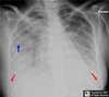
He has come in with a 2 week history of worsening shortness of breath, cough and hemoptysis. He was hypotensive on arrival with BP 80/40 and exam by the ER physician heard right sided crackles, so has been treated with ceftriaxone and azithromycin for CAP and given 2L fluid for septic shock.
However he has deteriorated and you are called down to assist in the ER and find this man tachypneic at 40, saturating 85% on 15L via non rebreather, a blood pressure of 75/50 and peripherally shut down. He is drowsy but able to obey commands.
His venous blood gas shows ph 7, lactate 9, BD 25. Hb 16, WBC/plts normal, INR 3, APTT 80, Cr 3, CRP 40.
What are your thoughts at this stage and how would you approach? I shall provide any more info requested.