Ok, I think I can answer this.
These researchers found that normal human cells have this protein, but cancer cells are deficient in it. So they want to know why, and they hypothesized that the mRNA produced by cancer cells is less stable than in normal cells. By less stable, they mean that it degrades more quickly (has a shorter half life). They think that it's because of a mutation they found on the 3'UTR. This one mutation on this UTR is the only difference on the mRNA between the cell types. To test this, they did two experiments. I think this is at least close to being right.
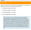
In the second experiment, they tested if the mutated 3'UTRs from the cancer cells had an effect on transcription compared to the WT. They put the 3'UTR from each of the cell lines on a luciferase reporter, and then measured bioluminescence 48 hours later. Transcription of mRNAs carrying the 3'UTRs from both cancer cell lines was way higher than transcription of the normal 3'UTR. This tells us that the protein deficiency is not because the mRNA is less transcriptionally active. But we conducted this experiment in Hela cells, could it be because the cancer cells themselves are less transcriptionally active? No, because the passage says that transcription of the gene proceeds normally. Lol. Also, just in general it's safe to assume that cancer cells are overall highly transcriptionally active. So it's not due to transcription.
So if the cancer cells are still making good levels of mRNA, why are they deficient in the protein? Could be that the mRNA is unstable and quickly gets degraded. So how do they measure this? From your findings in the luciferase experiment, you could predict that just doing straight up RT-qPCR would show you the same levels of transcript across all the cells. In fact, I bet that's exactly what they did to "verify that transcription proceeds normally."
So the solution is to block all transcription, wait 12 hours, and then do RT-QPCR. These cells haven't made any new transcripts in the last 12 hours. If we still see the same levels of this mRNA, that means that it's not really degrading, which means its stable. If we see a decrease in this mRNA, that means the mRNA is getting degraded and it's unstable.
This would have been a lot more straightforward if they had showed you a figure with two panels, one before the ActD and one 12 hours later. But they would like you to prove that you are worthy of a minty white coat through deductive guessing and decoding of intentionally badly written experimental design

 Screen Shot 2017-05-11 at 8.18.20 PM.png130.1 KB · Views: 62
 Screen Shot 2017-05-11 at 8.18.28 PM.png145.7 KB · Views: 61
 Screen Shot 2017-05-11 at 8.18.40 PM.png107.4 KB · Views: 46
 Screen Shot 2017-05-11 at 8.18.51 PM.png58.5 KB · Views: 56
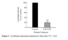 Screen Shot 2017-05-11 at 8.18.58 PM.png34.5 KB · Views: 62