Pictures of the Week

This forum made possible through the generous support of SDN members, donors, and sponsors. Thank you.

Epidural

Pain Moderator
Moderator Emeritus
Lifetime Donor
15+ Year Member
Joined
May 1, 2005
Messages
112
Reaction score
7
Each week we hope to add pictures to this thread for discussion. Please feel free to add pictures from your practice. Images need to be resized to 400 x 400. If you need help with uploading attachments, please send me a private message or e-mail us at [email protected]. Thank you.


Copyright © 2005 by PainRounds.com. All rights reserved. Terms of Use Policy.

Members don't see this ad.
 
During a transforaminal blunt needle block, the first picture depicts the vascular pattern obtained which clearly fills Batsons plexus and the contralateral extraspinal veins. The second picture is one second later. The third is with proper repositioning of the needle and the neurogram obtained in real time injection. Click on the pictures to enlarge.
 

Attachments

  • LRBwithVascularFlash.JPG
    LRBwithVascularFlash.JPG
    22.5 KB · Views: 3,655
  • LRBwithPostVascularFlash.JPG
    LRBwithPostVascularFlash.JPG
    20.7 KB · Views: 3,171
  • LRBwithPostRepositioningNeurogramRealTime.JPG
    LRBwithPostRepositioningNeurogramRealTime.JPG
    19.9 KB · Views: 3,152
Lack of knowledge regarding the course of the cervical medial branches can lead to misplaced RF needles and a failed procedure. In the case below, one lesion was created at each cervical lateral articular pillar. Unfortunately, the RF needle missed the medial branches as demonstrated in the right panel. It is important to understand the cervical medial branch anatomy before attempting RF. The anatomy may be viewed in the ISIS Guidelines section of this website. Click on the pictures to enlarge.
 

Attachments

  • CervRFwithoutNervesDrawnIn.JPG
    CervRFwithoutNervesDrawnIn.JPG
    13.7 KB · Views: 2,995
  • CervRFwithNervesDrawnIn.JPG
    CervRFwithNervesDrawnIn.JPG
    14.2 KB · Views: 2,921
Members don't see this ad :)
Selective nerve root blocks are so called due to the limited volume of anesthetic deposited into the neuroforamen with tracking to that single segment of the nerve in the cervical spine, or in the lumbar spine to the level of the disc herniation. Small sequential volumes of contrast are injected with fluoroscopy used in-between injections through a stationary needle. The volume required to achieve the goal of being selective is noted, then the same volume of local anesthetic is applied through the needle. Unfortunately, the physician below simply blasted the area with contrast with one injection thereby obliterating the volume data needed for selectivity of the local anesthetic application. The same volume of local anesthetic injected as seen below would eradicate any selectivity and potentially cause a false positive block due to local anesthetic spread over several nerves.
 

Attachments

  • NonSelectiveCervicalNRB.JPG
    NonSelectiveCervicalNRB.JPG
    24.3 KB · Views: 2,439
This unfortunate patient developed an enlarged pump pocket with intrathecal pump freely rotating each day after the fascial stitches ruptured. She came into my practice after this had been occurring for over a year and complained it was increasingly difficult to fill her Codman pump because of the rotation. Also there was a significant edema and fluid collection prior to each refill. For the past year the fluid had been drained (usually 100-150cc) in order to fill the pump. On our evaluation, the patient did indeed have a rotating pump and had 100cc non-colored clear fluid collecting under the skin. Elective surgery was undertaken to fix the pump to the fascia and the picture below was encountered. The catheter had over 30 loops in it due to the repetitive rotations of the pump and a significant amount of clear fluid was drained from around the pump. The catheter was also frayed and slighly split near the metal catheter connector. The Flex tip portion was trimmed, the catheter from the pump was trimmed, and a new catheter connector was used. Interestingly it was not possible to disconnect the Flex tip catheter from the catheter connector even using hemostats (frozen screw threads). It was decided the fluid collection had been a CSF hygroma from continuous leakage from the slight split in the catheter. The pump was sutured to the fascia with size 0 Ethibond stiches, the pump pocket imbricated, and closed. She had no more fluid collections in the future and the pump has remained fixed onto the fascia. Contributed by AlgosDoc.
 

Attachments

  • Torqued Catheter.JPG
    Torqued Catheter.JPG
    39.4 KB · Views: 2,532
algos,

nice case

did you infer a CSF hygroma since the catheter slit was distal to all the knots...implying the only fluid that could leak would be CSF? or did you test it, e.g for Beta Transferrin or mix it with pentobarbital (intra-operatively)


..we had a similar problem with the medtronic pump, but ours developed in a pump that had been fine for over 2 years, but then developed over the course of 2-3 follow-up visits......curiously, records showed that the pump refill went fine on the first visit...but by the second visit, some fluid had accumulated and the pump was very hypermobile; we were obligated to exclude a CSF hygroma, but I doubted it on clinical grounds...the patient had no neurological signs...e.g. spont intracranial hypotension or subdural hemmorhage..head CT was negative

also, the rate of CSF production (>500cc/day) accumulation of fluid would have been substantially higher than our bedside aspirate (about aobut 30-40cc) and the fluid analysis was negative for beta transferrin.. we were able to successfully refill the pump; notably he reported that his pain relief had deteriorated over the past month

by the third visit the patient's pump had flipped, but we refilled it successfully....he stated that he had increased yawning and GI motility and anxiety over the past few weeks (since the second visit)...not much diaphoresis

we took him to the OR and sure enough the pump and catheter were twisted....but not as tightly as yours...but curiously there were multiple holes in the catheter...this segment was removed and replaced.

I believe that the catheter was punctured during one of the refills...the patient was getting opioids, by default, parenterally rather than intrathecally..however the leaked fluid caused hydrodissection and released the pump and the non-absorbable retaining sutures (prolene)....

the pump then flipped...during the second refill, our 'flipping back' the pump...may have partially twisted the catheter...so that the patient got less parenteral drug (partially sealing off fluid leak into the pocket)...mild withdrawal

hence...even refilling flipped pumps can be hazardous!
 
I have had a couple of these flipped pumps. They seem to be in obese people, and they report that when they sit on the toilet they can feel the pump flip. I think the rolls of fat turn the pump. Maybe we should put these in the lower abdomen for obese people. Or maybe the buttock - there's lots of room.
 
Cervical intra-articular facet injections may be useful if there is insufficient clinical grounds (based on medial branch block response) to proceed to radiofrequency neurotomy. Using a caudad beam rotation of about 30 degrees to the cervical spine, one can often visualize the joints directly. Because the volume accommodated by the cervical z-joints is very small (often 0.25-0.5ml), mixing local anesthetics with steroids may exceed this volume for injection and can spill over into the epidural space, causing some nerve root anesthesia. Using small judicious volumes of injectate can also avoid overdistension of the joints. Contributed by AlgosDoc.
 

Attachments

  • CervIAFacetPosteriorApproach.JPG
    CervIAFacetPosteriorApproach.JPG
    17.6 KB · Views: 2,321
nice pictures
 
  • Like
Reactions: 1 user
Hello Epidural,

I found this fascinating MRI of a degenerated disc and thought you might consider it for picture of the week. :laugh:

No, it is not Siamese twins.
 

Attachments

  • DegenDisc2.jpg
    DegenDisc2.jpg
    47.9 KB · Views: 3,292
spinepain said:
Hello Epidural,

I found this fascinating MRI of a degenerated disc and thought you might consider it for picture of the week. :laugh:

No, it is not Siamese twins.

Lots of porn on the internet, but I do not think this qualifies.
 
greywater said:
I have had a couple of these flipped pumps. They seem to be in obese people, and they report that when they sit on the toilet they can feel the pump flip. I think the rolls of fat turn the pump. Maybe we should put these in the lower abdomen for obese people. Or maybe the buttock - there's lots of room.


As a potential patient and new home for a pump, and being over wieght since injuring my back, why not in the arm pit? or in the rump? Most large people sleep on their sides, so just keep it in the back??

Is there any doc's out there with these devices implanted? I would love to hear from you.
 
Members don't see this ad :)
Dr. Idrissi (fellow) and I put down some stereo cable. It was my idea to put three across at the same level. The patient had 722.83 as a prior Dx (L4-5 microdiskectomy) and failed all prior conservastive care before being referred for SCS. Axial>radicular pattern.

tripole.jpg
 
Just wanted to share a misadventure with kyphoplasty! Suprisingly the patient did well afterward and nothing was sticking out of her back!!! Lesson for the day- Twist and wiggle as you withdraw the kypho needle!!! Paravert--> I told you I would post it! hehe

B
 

Attachments

  • kyphoplasty gone wrong.jpg
    kyphoplasty gone wrong.jpg
    33.4 KB · Views: 1,926
kypholateral.jpg kyphomadness.jpg


So here's a typical case in the Lone Star State. This is how it's supposed to go. This is a 4 level kyphoplasty (myeloma), done under GA. It took me about 90 minutes of operative time, patient felt great the next day. These pictures are from the case that was done immediately prior to the pics posted above by my partner in crime.

So, here's the deal with the case above. We normally use the PMMA cement from Stryker regardless of whether we do kypho or vertebro, but the Kyphon rep wanted us to try his cement. Turns out, that stuff takes several more minutes to harden. In that case, as we were injecting through the left pedicle trocar, we noted some spread of cement into the posterior 1/3 of the vertebral body. We stopped injecting and waited about 4 minutes before we took the trocar out. As you can see, the cement hardened inside the trocar but did not break off at the pedicle like it is supposed to.

Either way, we told the patient about this immediately after the procedure. She did not complain of any focal tenderness at the site. She continues to deny any soft tissue pain in this area. In fact, she is very satisfied with her results. I suppose if it becomes a problem later, we can excise some of the more superficial cement. Unless it becomes a HUGE bother to her, I don't see any need to dissect down to the pedicle to get it all out.
 
Just wanted to share a misadventure with kyphoplasty! Suprisingly the patient did well afterward and nothing was sticking out of her back!!! Lesson for the day- Twist and wiggle as you withdraw the kypho needle!!! Paravert--> I told you I would post it! hehe

B


I'm jealous...Looks fun...I want to play with cement!
 
Here are some really good quality fluoro shots from a Gasserian I did a couple of weeks ago. Here's a tip I picked up to help get good lateral images -- look carefully at the teeth and try to align the shadows. I did it in this pic, and the rest of the anatomy is really clear.

I liked it so much, I made it my avitar.
 

Attachments

  • gasserian3.jpg
    gasserian3.jpg
    19.8 KB · Views: 1,574
  • gasserian2.jpg
    gasserian2.jpg
    18.4 KB · Views: 1,497
  • gasserian.jpg
    gasserian.jpg
    18.9 KB · Views: 1,639
OK, so here's the follow-up to my case that bbruel posted on my "behalf". So, the patient came to clinic two weeks later pissed off because it felt like someone was stabbing her in the back whenever she lied down. We figured that she had about a 3cm spike of hardened PMMA from the subcutaneous fat, extending through the muscles down to the facet joint. No problem.

So here's what I did. First off, I infiltrated the surrounding tissues with 10cc Lido 1% with epi. Then, we used fluoro to "gunbarrel" the spike (oblique.jpg).

Next, I made a 3cm incision parallel to the spine over the distal tip of the cement and blunt dissected down to lumbodorsal fascia. I used a Bovie, going around the cement spike to create a channel around it. Then I used a long, heavy duty clamp to nip the spike off at the base and pulled the thing out (lateral1.jpg).

The defect going down through the muscles to the facet was just big enough to get my index finger into. There was still about 1cm of PMMA remaining, so I nipped the rest out with a small rongeur. You can see that the PMMA goes all the way through the pedicle and stops just at the posterior wall of the facet (lateral2.jpg).

I irrigated the wound and closed in layers. Here are some pictures of the cement. The first has each piece spaced out (PMMA1.jpg), then put back together as best I could to demonstrate total length (PMMA2.jpg).

I think I'll send this as a letter to the editor to show how easy this is to do. Although this problem is fairly rare, it is the most common complication that patients complain about. Good to know I can easily deal with it if it happens again, or if I see somebody else's.
 

Attachments

  • latxray.jpg
    latxray.jpg
    15 KB · Views: 1,155
  • oblique.jpg
    oblique.jpg
    19.4 KB · Views: 1,144
  • lateral1.jpg
    lateral1.jpg
    13.6 KB · Views: 1,097
  • PMMA2.jpg
    PMMA2.jpg
    52.4 KB · Views: 993
  • PMMA1.jpg
    PMMA1.jpg
    23.9 KB · Views: 984
  • lateral2.jpg
    lateral2.jpg
    15.6 KB · Views: 1,003
  • Image1.jpg
    Image1.jpg
    7.7 KB · Views: 1,028
I think this is an old technique which has some utility in extremely tight neuroforamina. Indicated when a standard transforaminal cannot be placed due to osteophytes. Use a 20ga introducer and a 25ga inner needle. Place a very aggresive curve on the 25ga as with discography such that the 25ga veers medially as it emerges from the introducer needle. Note the tip of the 25ga needle at the 6 o'clock position on the pedicle. This particular patient is 400lbs so image quality is not great. 5" 20ga introducer and 8" 25ga epidural needle.
 

Attachments

  • twoneedle.jpg
    twoneedle.jpg
    34.8 KB · Views: 1,270
Nothing exciting, just lumbar discogram with contrast extending into a Schmorl's node on AP and lateral views.
 

Attachments

  • IMAGE001_2.jpg
    IMAGE001_2.jpg
    33.7 KB · Views: 1,064
  • IMAGE002_2.jpg
    IMAGE002_2.jpg
    32.5 KB · Views: 1,013
I tried a transforaminal lateral recess block as instructed by algosdoc. This patient has a large right paracentral HNP at L4/5. L5 radic symptoms. Previous right L5/S1 TFESI did not show contrast tracking up to level of pathology. I wanted to wash out the inflammatory soup at the HNP so directed a 8" 20ga epimed BLUNT tip needle to make direct contact with the bulging annulus, and injected steroid and saline. Patient tolerated the procedure roughly the same as the prior TFESI and no complications. A/P, Oblique, and Lateral images. On the lateral image you can see the needle tip being deflected dorsally and caudally by the large annulus bulge which was later outlined by contrast.

You can find a better technical writeup at algosdoc's website:
http://algosresearch.org/Techniques/TFLRB.html
 

Attachments

  • lrbap.jpg
    lrbap.jpg
    42.6 KB · Views: 1,376
  • lrblat.jpg
    lrblat.jpg
    31.8 KB · Views: 1,361
  • lrboblique.jpg
    lrboblique.jpg
    35.6 KB · Views: 1,347
What makes you think that a 20 gauge needle can be pushed by HNP?
 
I thought I'd try an resurrect this sticky. This is a simple case, but hopefully helpful to those in training and a reminder to the rest of us.

Saw this today - x-ray and MRI of L-Spine with a calcified mass in the right lower lumbar soft tissue. On AP it's clearly to the right of the spine, on lateral it is posterior, but the MRI shows that it is SubQ and not in the muscle.

This most likely represents a calcified granuloma from an old TPI - and the needle likely did not get into the muscle, but remained in the adipose tissue.

Caveat - when doing TPI's and IM injections, make sure your needle is long enough, 1.5" won't do it on many Americans these days.
 

Attachments

  • MRI axial.jpg
    MRI axial.jpg
    34.7 KB · Views: 964
  • AP.jpg
    AP.jpg
    23.2 KB · Views: 996
  • lat.jpg
    lat.jpg
    14.8 KB · Views: 900
I thought I'd try an resurrect this sticky. This is a simple case, but hopefully helpful to those in training and a reminder to the rest of us.

Saw this today - x-ray and MRI of L-Spine with a calcified mass in the right lower lumbar soft tissue. On AP it's clearly to the right of the spine, on lateral it is posterior, but the MRI shows that it is SubQ and not in the muscle.

This most likely represents a calcified granuloma from an old TPI - and the needle likely did not get into the muscle, but remained in the adipose tissue.

Caveat - when doing TPI's and IM injections, make sure your needle is long enough, 1.5" won't do it on many Americans these days.

also make sure you dont use corticosteroids.....
 
OK, so here's the follow-up to my case that bbruel posted on my "behalf". So, the patient came to clinic two weeks later pissed off because it felt like someone was stabbing her in the back whenever she lied down. We figured that she had about a 3cm spike of hardened PMMA from the subcutaneous fat, extending through the muscles down to the facet joint. No problem.

So here's what I did. First off, I infiltrated the surrounding tissues with 10cc Lido 1% with epi. Then, we used fluoro to "gunbarrel" the spike (oblique.jpg).

Next, I made a 3cm incision parallel to the spine over the distal tip of the cement and blunt dissected down to lumbodorsal fascia. I used a Bovie, going around the cement spike to create a channel around it. Then I used a long, heavy duty clamp to nip the spike off at the base and pulled the thing out (lateral1.jpg).

The defect going down through the muscles to the facet was just big enough to get my index finger into. There was still about 1cm of PMMA remaining, so I nipped the rest out with a small rongeur. You can see that the PMMA goes all the way through the pedicle and stops just at the posterior wall of the facet (lateral2.jpg).

I irrigated the wound and closed in layers. Here are some pictures of the cement. The first has each piece spaced out (PMMA1.jpg), then put back together as best I could to demonstrate total length (PMMA2.jpg).

I think I'll send this as a letter to the editor to show how easy this is to do. Although this problem is fairly rare, it is the most common complication that patients complain about. Good to know I can easily deal with it if it happens again, or if I see somebody else's.

I broke off a straw on a V-plasty 2 weeks ago. From pedicle to superficial in the paraspinals. I opened with the 11 blade 2cm, recannulated the cement into the V-plasty needle using a mosquito and my finger to guide it in. Took the needle down to the pedicle and applied sheer force inferolaterally. Broke the straw of PMMA at the pedicle and it came out with the needle. Saved it on my desk to remind me to replace the stylet before removing the needle.
 
missed.jpg


missed2.jpg


Blind epidurals. This was the first of 2. This one didn't work. The second one was a wet tap requiring blood patch. The patient came to me 2 months after these epidurals and was needle phobic. The MRI was obtained 9 months after the 1st ESI.

(If you don't read MRI well, look behind the spinous process at L4 in the subQ fat) In the sag image, I measured 3.3cm from the placement of the prior ESI to the epidural space. Using US to drain out the fluid this afternoon.

This would be a great case report, great visuals.
 
59 y/o WF with chronic LBP radiating down left leg postrior thigh and calf as aching, burning, stabbing, 8/10. No weakness, no sensory loss, normal DTR at ankle, not tested at knee because of recent arthroscopic knee surgery. MRI x2 with mild L4-5 spondylosis without foraminal or canal stenosis, no disc HNP, bone island at S2. Seen by NS, OSS, and 2 pain clinics before me. Failed ESI's as TF and IL approaches. Had some relief with SIJ/Piri injection but short lived. Multiple opioids tried, multiple neuropathic agents used.

Still no relief. Bone scan for sacral insuff Fx negative. Labs negative.
Sees 3rd opinion NS in another state. He feels weak pulses in the foot, asks for vascular studies.


iliac.jpg


Left main iliac disease mimics L5/S1 radicular pain.

Vascular surgery consulted. Appt in 2 weeks.
 
The prevalence of occult peripheral arterial disease among patients referred for orthopedic evaluation of leg pain
Joseph Bernstein
Department of Orthopedic Surgery, University of Pennsylvania, Philadelphia, PA, USA; Veterans Affairs Medical Center, Philadelphia, PA, USA

John L Esterhai

Department of Orthopedic Surgery, University of Pennsylvania, Philadelphia, PA, USA; Veterans Affairs Medical Center, Philadelphia, PA, USA

Mitchell Staska

Veterans Affairs Medical Center, Philadelphia, PA, USA

Sally Reinhardt

Veterans Affairs Medical Center, Philadelphia, PA, USA

Marc E Mitchell

Department of Surgery, University of Mississippi, Jackson, MS, USA [email protected]

Abstract

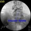
Lower extremity peripheral arterial disease (PAD) and musculoskeletal conditions both produce symptoms of leg pain, and may coexist. This study assesses the prevalence of PAD among patients referred to orthopedic surgery for evaluation of lower extremity pain. Fifty consecutive patients aged 50 years or more who had a chief complaint of leg pain, no history of trauma, and no previous history of PAD were studied prospectively. The presence of known risk factors for PAD and classic claudication symptoms was assessed by telephone interview and medical record review. Individuals were then evaluated by measurement of the ankle–brachial index (ABI) using Doppler and pulse volume recordings (PVR). A patient was deemed to have PAD if the ABI was below 0.9 or if the PVR demonstrated significant abnormalities. Occult PAD was detected in 10 of the 50 patients (20%) on the basis of the non-invasive vascular studies. There were no differences between patients with PAD and those without PAD regarding the presence of risk factors for PAD. None of the patients without PAD had claudication, while only one of the 10 patients with PAD had symptoms of classic claudication. In conclusion, 20% of patients referred by primary care providers to the orthopedic surgery clinic for lower extremity pain were discovered to have occult PAD. The majority of these patients did not have claudication. Orthopedic surgeons and primary care providers must maintain an appropriately high index of suspicion for PAD when evaluating patients with non-traumatic lower extremity pain.

Article out 1 month too late.
 
Just wanted to share a misadventure with kyphoplasty! Suprisingly the patient did well afterward and nothing was sticking out of her back!!! Lesson for the day- Twist and wiggle as you withdraw the kypho needle!!! Paravert--> I told you I would post it! hehe

B


oh........
 
Here are some really good quality fluoro shots from a Gasserian I did a couple of weeks ago. Here's a tip I picked up to help get good lateral images -- look carefully at the teeth and try to align the shadows. I did it in this pic, and the rest of the anatomy is really clear.

I liked it so much, I made it my avitar.


man, he has skills.......
 
just now decided to look through this sticky. LOVE it!!! great learning. post more if ya'll got em.
 
Picture of a Neurotherm Simplicity probe during sacral RFL.
 

Attachments

  • sacral RFL 351x320.jpg
    sacral RFL 351x320.jpg
    14.9 KB · Views: 797
This came up in a discussion of thoracic RF on the ISIS forum. After reviewing thoracic MB anatomy a while back I decided to place my probe over the pedicle shadow. The thoracic MB runs right across the area.

You have to come in at a steep angle so you can place the probe along the dorsal surface of the lamina. I enter over the pedicle shadow of the level below and target the inferior aspect of the pedicle shadow below the joint. Come in paramedian so you can come across the pedicle perpendicular to the nerve. I suppose it would be better if you were parallel to the nerve but then you'd have to enter from the contralateral side. I am just grateful to get a decent burn in this area and this gives me the best shot at catching the nerve.

For example, if I want to burn at T4 I enter at the pedicle shadow of T5. Then I aim for the pedicle shadow of T4. Sometimes you have to enter even more caudally, between the T5 and T6 pedicle shadows. I fish around for a sensory paresthesia and then do a 90 sec 80C burn.

To my surprise, this has worked very well. Here is a picture of two burns with the MBs drawn in. The circles under the tips of the probes are the pedicles and the yellow lines are the nerve pathways.
 

Attachments

  • tmB RF.jpg
    tmB RF.jpg
    6 KB · Views: 898
This came up in a discussion of thoracic RF on the ISIS forum. After reviewing thoracic MB anatomy a while back I decided to place my probe over the pedicle shadow. The thoracic MB runs right across the area.

You have to come in at a steep angle so you can place the probe along the dorsal surface of the lamina. I enter over the pedicle shadow of the level below and target the inferior aspect of the pedicle shadow below the joint. Come in paramedian so you can come across the pedicle perpendicular to the nerve. I suppose it would be better if you were parallel to the nerve but then you'd have to enter from the contralateral side. I am just grateful to get a decent burn in this area and this gives me the best shot at catching the nerve.

For example, if I want to burn at T4 I enter at the pedicle shadow of T5. Then I aim for the pedicle shadow of T4. Sometimes you have to enter even more caudally, between the T5 and T6 pedicle shadows. I fish around for a sensory paresthesia and then do a 90 sec 80C burn.

To my surprise, this has worked very well. Here is a picture of two burns with the MBs drawn in. The circles under the tips of the probes are the pedicles and the yellow lines are the nerve pathways.

The way I have seen it done is to go to the superolateral border of the transverse process - so transverse process/rib junction. I think because of this study?

Acta Neurochir (Wien) (1995) 136:140-144
W. H. Chua1 and N. Bogduk1

(1) Faculty of Medicine and Health Sciences, The University of Newcastle, New South Wales, Australia


Summary Thoracic percutaneous facet denervation has been employed for the treatment of thoracic zygapophysial joint pain. But the surgical anatomy of this procedure has been assumed to be the same as for lumbar medial branch neurotomy. To establish the anatomical basis for thoracic medial branch neurotomy, an anatomical study was undertaken. Using an X40 dissecting microscope, a total of 84 medial branches from 7 sides of 4 embalmed human adult cadavers were studied.
The medial branches of the thoracic dorsal rami were found to assume a reasonably constant course. Upon leaving the intertransverse space, they typically crossed the superolateral corners of the transverse processes and then passed medially and inferiorly across the posterior surfaces of the transverse processes before ramifying into the multifidus muscles. Exceptions to this pattern occurred at mid-thoracic levels (T5–T8). Although the curved course remained essentially the same, the inflection occurred at a point superior to the superolateral corner of the transverse process.
At no time during the dissection were nerves encountered crossing the junctions between the superior articular processes and transverse processes which have been the target points advocated for thoracic facet denervation. Rather, the results of this study indicate that the superolateral corners of the transverse processes are more accurate target points.

Keywords Thoracic nerves - dorsal rami - facet denervation - zygapophysial joint
 
I do the same as Gorback but come medial to lateral, caudal to rostral- in an effort to get more of the 10mm tip along the nerve as it wraps into the joint.

I have looked at Chua's work and it is one of the only published anatomical articles on thoracic MBB's. There was some 1990's stuff from Stolker or some name similar to that.

Also Stanton-Hicks 2004 article on Thoracic RF procedures:

History, Physics, and Clinical Proceures

RFA of Thoracic Medial Branch Nerves

Anatomy
Unlike the medial branch nerves in the lumbar region that pass
superiorly and medial close to the pedicle before passing under
the mammilo-accessory ligament and climbing up the articular
pillar superior to arborize on the capsule of the zygapophyseal
joint capsule, the thoracic medial branch nerves follow slightly
different courses between T1-T4, T5-T8, and T9-T10. At T11
and T12 each nerve follows a similar course to that in the
lumbar region (Chua and Bogduk,18 Chua,19 and Stolker and
coworkers20). Between T1 and T2, the medial branch nerves
cross the transverse process at the junction of the pedicle and
transverse process, passing medially and downward toward the
posterior surface of the transverse process. Between T5 and T8,
the medial branch nerve passes slightly superior and may not be
in contact with the transverse process. Between T9 and T10, the
nerve, after crossing the transverse process, may pass down
over the posterior surface of the transverse process.
 
gorback

what is your feedback on SImplicity (from Neurotherm) - i saw the picture you posted.

i have done the scorched earth technique for SI joints and have been unimpressed w/ results, and suspect this may be more of the same but with a cooler/easier/quicker way of doing it.

my neurotherm rep is hoping that i can start doing these - btw, they are quoting $500-600 per probe

your thoughts?
 
This was my first one and all I can say so far is that the postop pain is phenomenal. I have confirmed this with others. Bill Rittman at Neurotherm thinks it's due to the large burn, but the probe has a very sharp point and I think at least some of the pain might be mechanical trauma from scraping along the periosteum. I have encouraged him to try making a blunt-tipped probe and see if the postop pain is less.

Some of my colleagues have been enthusiastic. There seems to be a significant divide between those who think SI RF is wonderful and others who can't seem to make it work. The literature so far supports the latter conclusion.
 
do you think it was a patient-selection issue?

how long did you burn for - at what temperature? neurotherm rep was saying 5-8 minutes... i would prefer 2 minutes......

primarily, because the longer you char tissue the higher the incidence of infection....

now, if there was a delicate way of doing this both posteriorly AND anteriorly then it would be REALLY cool - if you don't puncture the rectum or cause massive pelvic floor dysfunction/laxity :(
 
This woman clearly has severe scoliosis and DJD. She had severe tenderness to palpation directly over the L5 transverse process batwing deformity/pseudoarticulation as confirmed by fluroscopy. I then proceeded to inject this area with a surprisingly nice arthrogram! I'm sure I'm not the first to ever do this, but I've never seen a published case nor a published arthrogram, so here is one for future reference. Might do a case report on it.
 

Attachments

  • 1.jpg
    1.jpg
    19.2 KB · Views: 1,042
  • 2.jpg
    2.jpg
    22.4 KB · Views: 942
  • 3.jpg
    3.jpg
    22.3 KB · Views: 906
I've been evaluating different needles for TFESIs. Today, I tried out a 29 gauge 3.5" Sprotte tip needle from Pajunk, Germany. Very nicely made needle. It was so flimsy it could not penetrate the skin. Used an 18 gauge to do that. However, once through the skin, it "drove" like a F1 car. It pierced the intertransverse ligament without difficulty, which I was surprised about. I'm on the lookout for 31 gauge spinal needles now!
 

Attachments

  • 1.jpg
    1.jpg
    21.9 KB · Views: 821
Any suggestions on non-surgical treatment for the bat-wing deformity. I have a patient with fusion on the left, not right, and mild-moderate facet sclerosis at L4-5 and L5-S1. Symptoms more consistent with sacroiliitis. No response to MBB or to SI injection.
 
Any suggestions on non-surgical treatment for the bat-wing deformity. I have a patient with fusion on the left, not right, and mild-moderate facet sclerosis at L4-5 and L5-S1. Symptoms more consistent with sacroiliitis. No response to MBB or to SI injection.

Did you see my images two posts above? you could try an injection like that and see if it helps.
 
I placed the needle in a similar position and contrast tracked all the way back to within 5 mm of the L5 vertebral body. Her articulation extended up on to the ilium as if to extend her SI joint and so I injected that extension as well. She got off the table with a smile on her face. We'll see. Thanks.
 
Saw this today. 59 yo with polio as a child.

His lower C-Spine is actually parallel to the ground.

Pt came to see me for lower back back. Just wants an NSAID. Does his own HEP.

Wish I could load original size, hopefully you can enlarge it.
 

Attachments

  • scoli2.jpg
    scoli2.jpg
    13.2 KB · Views: 812
Top